Request Sent a Appointment
Services
Colour Doppler
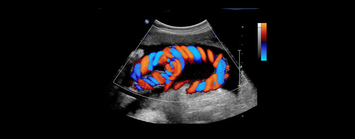
Color Doppler is an advanced ultrasound imaging technique that helps visualize the movement of blood within vessels and the heart by using sound waves. Unlike standard ultrasound, which only shows the structure of organs and tissues, Color Doppler adds information about the direction, speed, and quality of blood flow. It displays blood flow in different colors (commonly red and blue), where each color indicates whether blood is flowing towards or away from the ultrasound probe. This makes it a valuable tool in diagnosing vascular conditions, detecting blockages, and evaluating blood circulation in real time.
Detecting vascular blockages or narrowing (arterial stenosis, venous thrombosis)
Evaluating heart function and detecting abnormal blood flow patterns (valvular defects, congenital heart disease)
Assessing pregnancy for monitoring blood flow to the placenta and fetus
Checking organ perfusion (liver, kidneys, etc.)
Monitoring post-surgical vascular grafts or stents
Procedure
A water-based gel is applied to the skin to transmit sound waves efficiently.
A handheld device called a transducer is moved over the target area.
The ultrasound machine uses the Doppler effect to measure changes in frequency of sound waves reflected by moving blood cells.
Results are displayed as a color map overlay on the grayscale ultrasound image (usually red/blue).
Types of Doppler Ultrasound
Color Doppler – Shows blood flow direction and speed in color.
Power Doppler – More sensitive, shows blood flow even in very small vessels, but without direction.
Spectral Doppler (Pulsed/Continuous) – Provides a graph showing detailed flow velocity over time.
Duplex Doppler – Combines grayscale imaging with Doppler for anatomical + flow assessment.
Advantages
Non-invasive and safe (no radiation).
Provides real-time assessment.
Detects early vascular abnormalities.
Useful for pregnant women and children.