Request Sent a Appointment
Services
3D/4D/5D USG Sonography
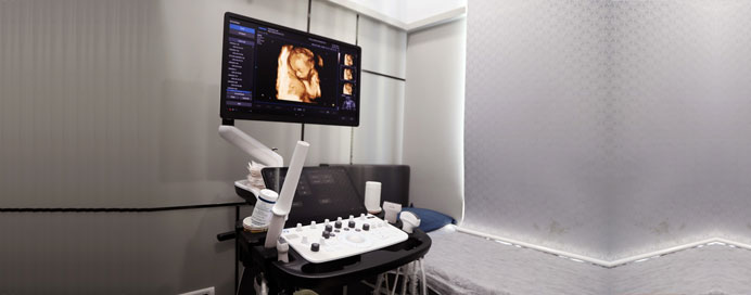
USG (Ultrasonography) / Sonography is a safe, non-invasive imaging technique that uses high-frequency sound waves to create images of the body’s internal organs and tissues. Over the years, ultrasound technology has advanced beyond traditional 2D imaging into 3D, 4D, and now 5D sonography, providing doctors and patients with highly detailed and dynamic views. These advanced techniques are especially valuable in obstetrics, gynecology, cardiology, abdominal studies, and musculoskeletal imaging.
3D Sonography
Definition: Produces three-dimensional still images by compiling multiple two-dimensional scans.
Uses: Helps in evaluating the fetus’s anatomy, face, spine, limbs, and detecting congenital abnormalities more accurately than 2D ultrasound.
Advantages: Provides realistic visualization for better diagnosis and parental bonding during pregnancy.
4D Sonography
Definition: An advanced version of 3D ultrasound that shows real-time moving images (like a video).
Uses: Displays fetal movements, facial expressions, heartbeats, and even yawns or smiles inside the womb.
Advantages: Improves accuracy in detecting abnormalities, enhances parental experience, and assists doctors in evaluating fetal growth and development in real time.
5D Sonography
Definition: The latest technology that combines 3D/4D imaging with advanced software enhancements for crystal-clear, high-definition images with better depth and contrast.
Uses: Provides superior visualization of fetal structures, organs, blood flow, and overall growth.
Advantages: Offers lifelike, high-definition images that aid in more precise diagnosis, especially for high-risk pregnancies.
Applications of 3D/4D/5D Sonography
Obstetrics – Monitoring fetal development, identifying birth defects, and evaluating placenta and amniotic fluid.
Gynecology – Assessing uterus, ovaries, and reproductive organs.
Cardiology – Visualizing heart structures and blood flow.
Abdominal Imaging – Detecting liver, kidney, gallbladder, and pancreatic disorders.
Musculoskeletal – Examining joints, ligaments, and muscles.
Benefits
Safe, non-invasive, radiation-free imaging.
More accurate detection of abnormalities.
Realistic, clear images for better understanding.
Enhances doctor-patient communication.
Strengthens parental bonding with the unborn baby.
Sonography & Ultrasound Studies
- Routine Sonography
- Obstetrics Sonography
- 5D Sonography
- Colour Doppler Studies
- Liver Elastography
- 2D Echocardiography
- Foetus Echocardiography
- Musculoskeletal Sonography
- Transrectal Sonography
- Transvaginal Sonography
- TIFFA Scan / Fetal Anomaly Scan
- NT (Nuchal Translucency) Scan
- Fetal ECHO
- Fetal Growth Scan